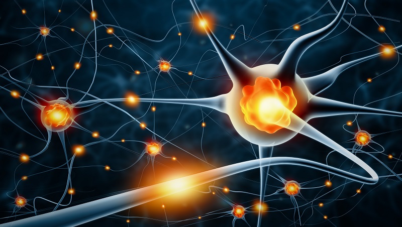

The neuron is a small information processor, and dendrites serve as input sites where signals are received from other neurons. The soma has branching extensions known as dendrites. The nucleus of the neuron is located in the soma, or cell body. This illustration shows a prototypical neuron, which is being myelinated by a glial cell. This membrane allows smaller molecules and molecules without an electrical charge to pass through it, while stopping larger or highly charged molecules.įigure 1. A neuron’s outer surface is made up of a semipermeable membrane. Like all cells, neurons consist of several different parts, each serving a specialized function. Neurons are the central building blocks of the nervous system, 86 billion strong at birth. This section briefly describes the structure and function of neurons. Neurons, on the other hand, serve as interconnected information processors that are essential for all of the tasks of the nervous system. This is important because it suggests that human brains are more similar to other primate brains than previously thought (Azevedo et al, 2009 Hercaulano-Houzel, 2012 Herculano-Houzel, 2009). For years, researchers believed that there were many more glial cells than neurons however, more recent work from Suzanna Herculano-Houzel’s laboratory has called this long-standing assumption into question and has provided important evidence that there may be a nearly 1:1 ratio of glia cells to neurons. Glial cells provide scaffolding on which the nervous system is built, help neurons line up closely with each other to allow neuronal communication, provide insulation to neurons, transport nutrients and waste products, and mediate immune responses. Glial cells are traditionally thought to play a supportive role to neurons, both physically and metabolically. The nervous system is composed of two basic cell types: glial cells (also known as glia) and neurons. Learning how the body’s cells and organs function can help us understand the biological basis of human psychology. Psychologists striving to understand the human mind may study the nervous system. Explain the role and function of the basic structures of a neuron.The cookie is set by the GDPR Cookie Consent plugin and is used to store whether or not user has consented to the use of cookies. The cookie is used to store the user consent for the cookies in the category "Performance". This cookie is set by GDPR Cookie Consent plugin. The cookie is used to store the user consent for the cookies in the category "Other. The cookies is used to store the user consent for the cookies in the category "Necessary". The cookie is set by GDPR cookie consent to record the user consent for the cookies in the category "Functional". The cookie is used to store the user consent for the cookies in the category "Analytics". These cookies ensure basic functionalities and security features of the website, anonymously. Necessary cookies are absolutely essential for the website to function properly. There are not such vesicles in the dendrites.Īxon contains neurofibrils all over but they lack Nissl’s granules.ĭendrites contain both neurofibrils and Nissl’s granules.Īxon conducts neuronal impulse away from the soma (cell body).ĭendrites conduct neuronal impulses towards the soma. No synaptic knots are formed on the tip of the dendrites. The terminal branches f the axon forms an enlarged synaptic knot. The thickness of axon is uniform throughout the length.ĭendrites are highly branched throughout their length. (Image Source: CC Wikipedia) You may also like NOTES in.Īxon arises from the discharging end of the nerve.ĭendrites arise from the receiving surface of the nerve.Īxon is comparatively long, sometimes several meters.ĭendrites are comparatively shorter (usually under 1.5 mm) Ø Both are cytoplasmic projections from the cell body of a neuronal cell. Ø Both axon and dendrites are the parts of neuron.


 0 kommentar(er)
